


Testicular cancer is the leading cancer in men ages 15-44, but can affect a man at any time in his life. Risk factors for developing testicular cancer are family history of testicular cancer, abnormal development of the testicles, and being of caucasian race. This month’s blog will be dedicated to the anatomy of the testicles, with a special focus on the draining lymph nodes, in honor of Testicular Cancer Awareness Month (April).
When abstracting a testicular cancer, you may have wondered about the location of the draining lymph nodes. The regional lymph nodes (Preaortic, Para-aortic, Paracaval, etc) are found next to the Aorta, just inferior to the kidneys. These lymph nodes are not at all close to the testes, which are found outside the abdominal cavity. The answer lies in the embryological development of the testes. The tissue that will become the gonads, testes in males, and ovaries in females, begins in the abdominal cavity (“ot” in Drawing A).
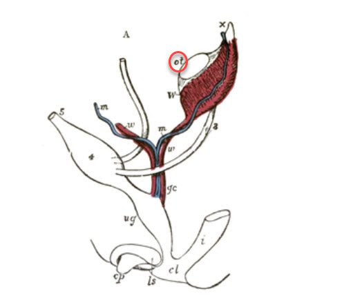
Images: By Henry Vandyke Carter – Henry Gray (1918) Anatomy of the Human Body Public Domain
In females, the tissue remains where it is, and develops into the ovaries (“o” in Drawing B).
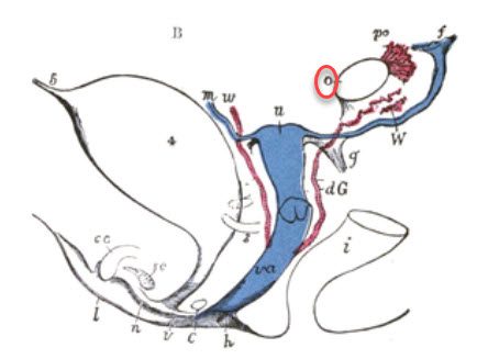
Images: By Henry Vandyke Carter – Henry Gray (1918) Anatomy of the Human Body Public Domain
In males, the testes will descend to the scrotal sac prior to birth (“t” in Drawing C).
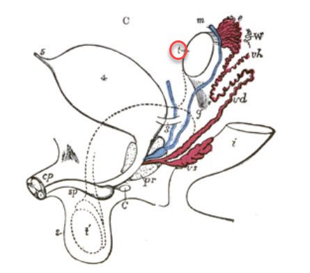
Images: By Henry Vandyke Carter – Henry Gray (1918) Anatomy of the Human Body Public Domain
The associated arteries, veins and lymphatics, likewise, descend to the scrotal sac. Lymphatics leaving the testes travel superior to lymph nodes next to the Aorta. This makes the external and internal iliac lymph nodes distant, even though they are physically closer to the testes.
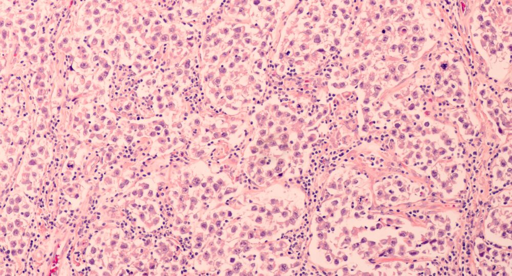
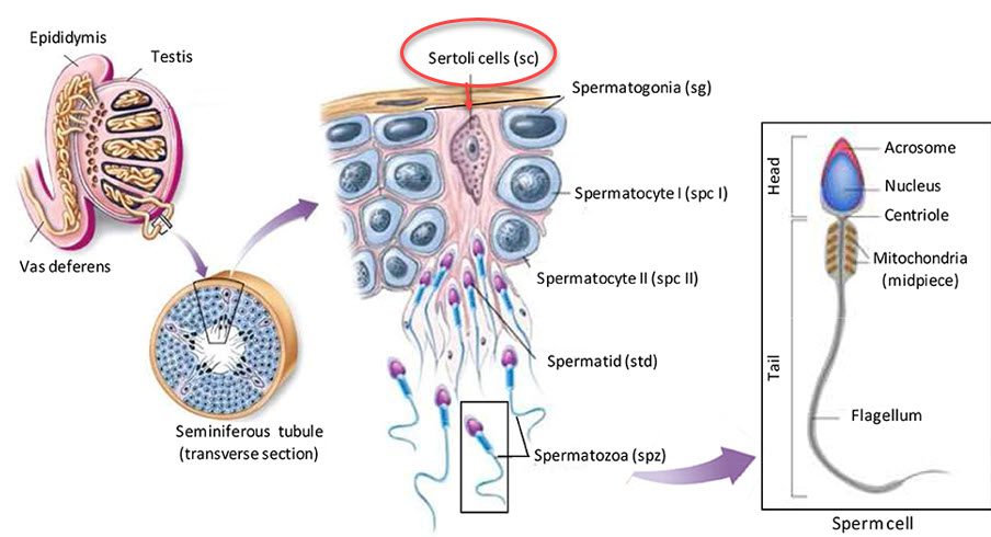
The testis contains several types of cells. The cells that develop into sperm are called Germ Cells. These cells are found within the seminiferous tubules. Also in the seminiferous tubules are Sertoli cells, which completely surround the germ cells. They help to support the germ cells and form the blood-testis barrier. Outside the seminiferous tubules are the Leydig cells. These cells make testosterone. Each of these cells can become cancerous.
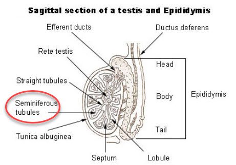
Image: National Cancer Institute: SEER Training
This Photo by Unknown Author is licensed under CC BY
Germ cell tumors are most common, with two main types: seminoma and non-seminomas; non-seminomas being choriocarcinoma, embryonal carcinoma, yolk sac tumor, and teratoma. Less common testicular cancers are Leydig cell tumor, Sertoli cell tumor, Carcinoma of the rete testis, and lymphoma.